Bumps and Blisters in the Mouth: Causes, Signs, and What to Do
Bumps and Blisters in the Mouth: Causes, Signs, and What to Do
Understanding how these fluid-filled swellings develop—and why attempting to pop them at home is never recommended—makes navigating professional oral care much simpler.
Understanding Common Oral Swellings: Mucocele vs. Ranula
Salivary glands throughout the mouth produce saliva through microscopic ducts. When one of these tiny pathways becomes pinched, injured, or blocked, fluid escapes into surrounding soft tissues rather than draining freely.
1. Oral Mucocele
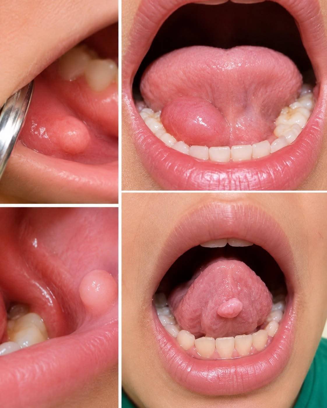
A mucocele is a harmless, mucus-filled cyst. It most commonly appears on the inner surface of the lower lip, though it can also form on the inner cheeks or the palate.
Typical Trigger: Often caused by mild, repeated micro-injuries, such as accidentally biting the inside of the lip or habit-based cheek chewing.
Appearance: A soft, domed nodule that appears translucent, bluish, or natural pink. It can range from a few millimeters to over a centimeter in width.
2. Ranula
A ranula is a specific, larger type of mucocele located on the floor of the mouth beneath the tongue.
Typical Trigger: Arises from a disruption in a major salivary gland (most often the sublingual gland).
Appearance: Larger, translucent, bubble-like swelling beneath the tongue that can sometimes push upward, making chewing, swallowing, or clear speech temporarily awkward.
Other Potential Causes of Oral Nodules
While mucoceles and ranulas account for many fluid-filled oral swellings, dental professionals also evaluate for other benign or chronic growths during an exam:
Irritation Fibromas: Firm, smooth nodules formed from fibrous connective tissue, typically caused by chronic friction against teeth or braces.
Squamous Papillomas: Small, wart-like growths on the mucosa.
Pyogenic Granulomas: Highly vascular, easily irritated bumps that occasionally bleed during brushing.
Salivary Gland Growths: Slower-growing nodules that require targeted clinical evaluation to rule out deeper gland issues.
Common Signs to Monitor
Most oral swellings do not cause intense pain unless they have been irritated by aggressive chewing. Watch for these everyday indications:
| Characteristic | What to Look For |
| Texture | Soft, smooth, compressible, or fluid-filled |
| Color | Clear, pale blue, or matching the natural pink of your gums |
| Sensation | Feeling like a tiny foreign object or smooth marble in the mouth |
| Functional Impact | Mild interference with chewing, smiling, or speaking |
| Fluctuation | The bump may occasionally rupture, shrink, and then refill over days |
Why You Should Never Pop an Oral Blister at Home
When a fluid-filled bump appears, the temptation to squeeze or pierce it with a needle is strong. However, home popping is generally counterproductive:
High Rate of Recurrence: Simply releasing the mucus leaves the damaged duct unaddressed. Once the outer layer seals, the swelling almost always returns.
Infection Risk: The oral cavity naturally hosts diverse bacteria. Introducing non-sterile tools increases the likelihood of secondary bacterial infections.
Tissue Scarring: Squeezing can tear surrounding tissue, turning a simple minor cyst into a more irritating chronic bump.
How Dentists and Oral Surgeons Address Them
If a mouth bump lasts longer than 10 to 14 days, consulting a general dentist or an oral and maxillofacial surgeon is the best path forward.
Clinical Examination: The provider evaluates the nodule’s shape, consistency, location, and history of accidental bites.
Targeted Imaging: Small in-office ultrasounds or soft-tissue evaluations may be used for deeper ranulas.
Minor In-Office Removal: A simple, quick procedure to remove the cyst and the affected minor gland prevents the pocket from refilling.
Marsupialization: For larger ranulas, a surgeon creates a small exterior opening with fine sutures to establish a continuous drainage path.
Active Monitoring: Small, asymptomatic mucoceles in children are sometimes monitored briefly, as some resolve spontaneously after minor trauma heals.
Disclaimer: This article is intended solely for educational and informational purposes and does not substitute for professional medical or dental advice, diagnosis, or clinical evaluation. Always consult a qualified dentist or oral surgeon if you notice persistent, growing, or uncomfortable oral lesions.